The Diary Game: 25.6.2026| How I spent my day in the histopathology laboratory.
A brief introduction
Hey! @steemit fam, good morning, good afternoon and good evening depending the time you’ll be reading this vlog, this is the period of my clinical posting of my academic career as a medical laboratory science student. Pls
stop bye to support this mini blog, if you love it give me a follow (follow the page) if you don’t still stick around till you see something that will make you follow this page❤️.
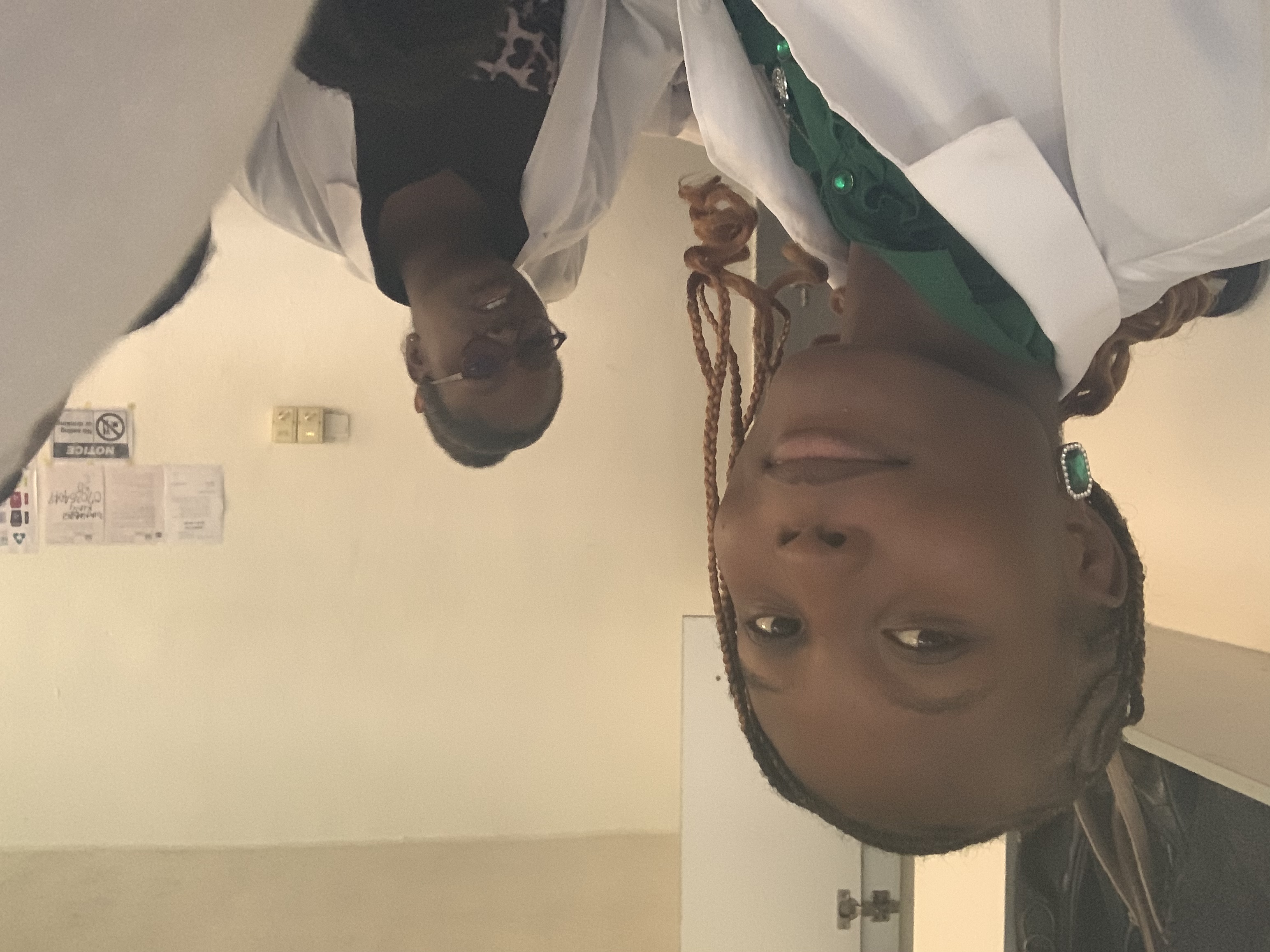
I and my colleague
General overview of the histopathology laboratory
The histopathology lab is a unique laboratory unit in the whole of medical laboratory science, it deals on the diagnoses of diseases in patients tissues. Samples used for diagnosis in histopathology are tissues, CSF fluid etc. This laboratory unit have two sub-unit, the histopathology and Cytology, the cytology comprises of non-gynecological(sample that are not from the reproductive organs)and gynecological(samples from the reproductive organ). So this different unit aid in the smooth examination of samples in this laboratory. It is therefore worthy to note that result interpretation are carried out by Pathologists.
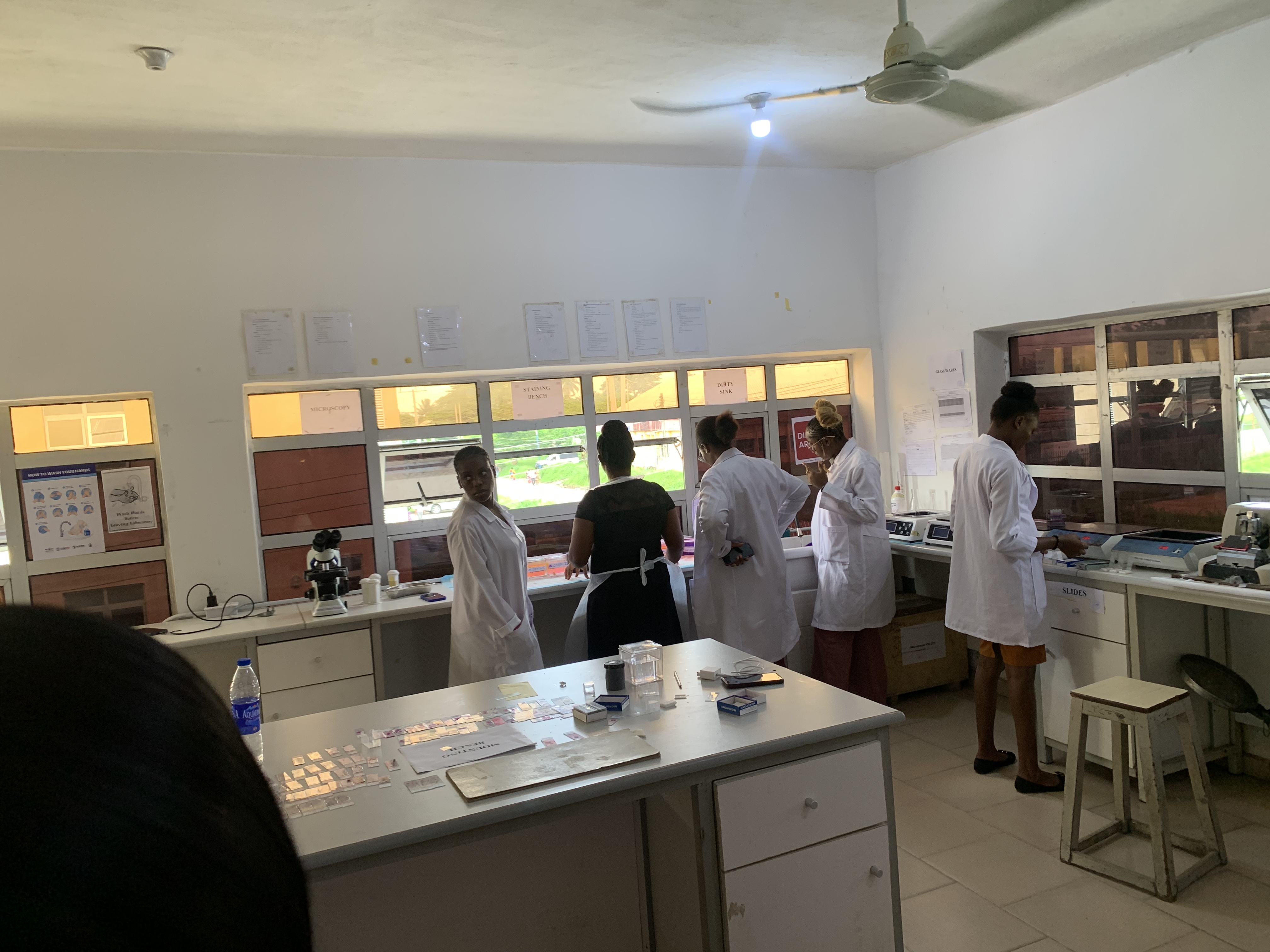
A unit of part of the laboratory
Apparatus In the laboratory
So they’re equipment that help in the successful processing of tissue. We have the automatic tissue processing, infiltration container, embedding machine, microtome, water bath, hot plate, and the staining section. So for the scientist to get a good and unique sample on a glass slide that will help in the visualization under a microscope and proper result interpretation it has to undergo various process with the above equipment listed. The tissue is being processed by the automatic tissue processor, then it’s been infiltrated in the infiltration container and then it’s been embedded with a paraffin wax, it’s sectioned on a microtome then section is place in water and picked up with a slide and then place on the hot plate in other for section to dry and adhere to the glass slide, then after few minutes it’s taken to the staining section for the sample to be stained and after going through the staining process, glass-slide is being mounted with a mountant and cover slipped to enable visualization, Note that this tissue sample is being sectioned and put in a sample cassette before the the above process occurs, and it’s for histopathological sample. I love you, stick around and let’s get connected. Sending loves ❤️
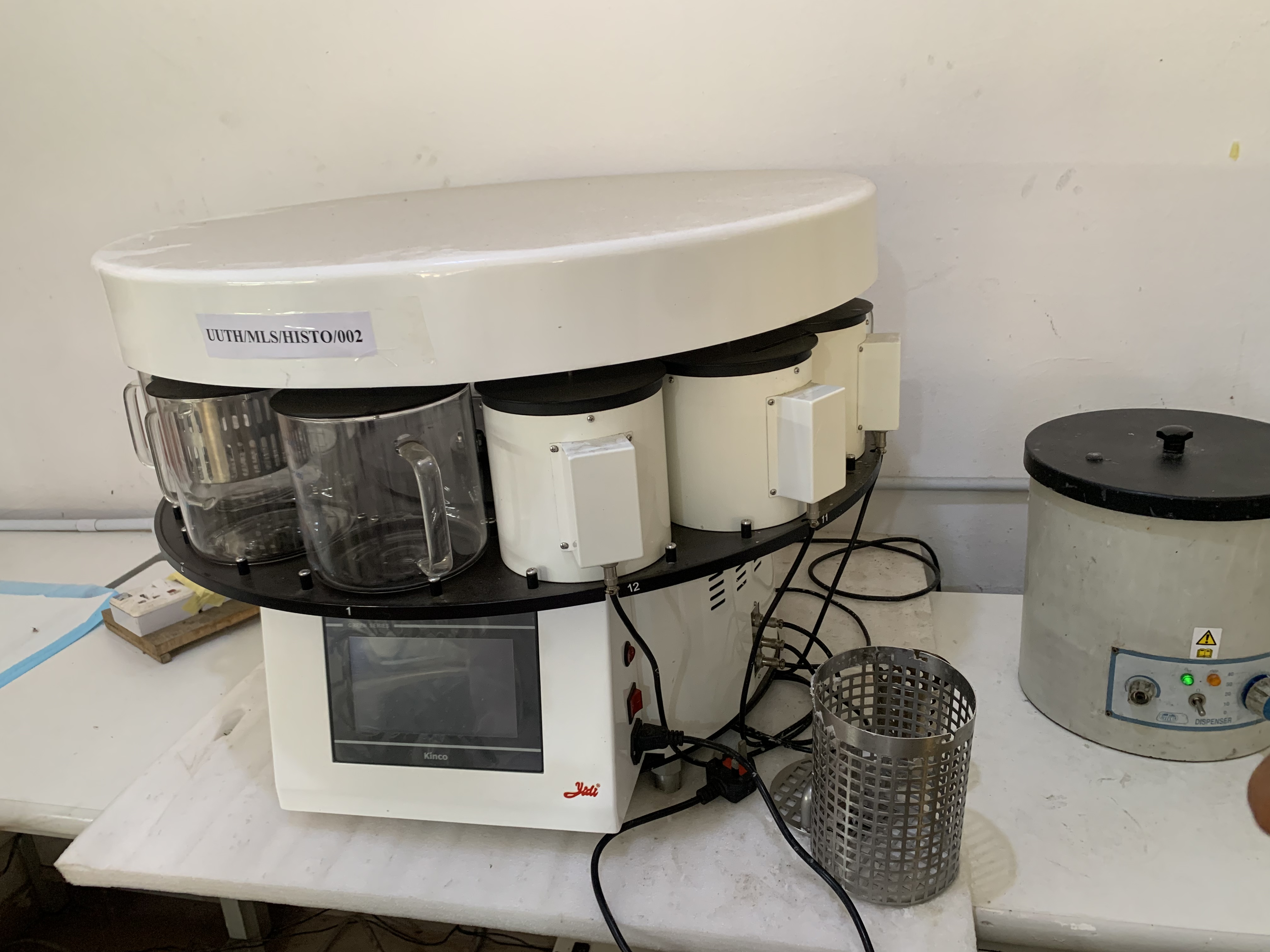
Tissue processor
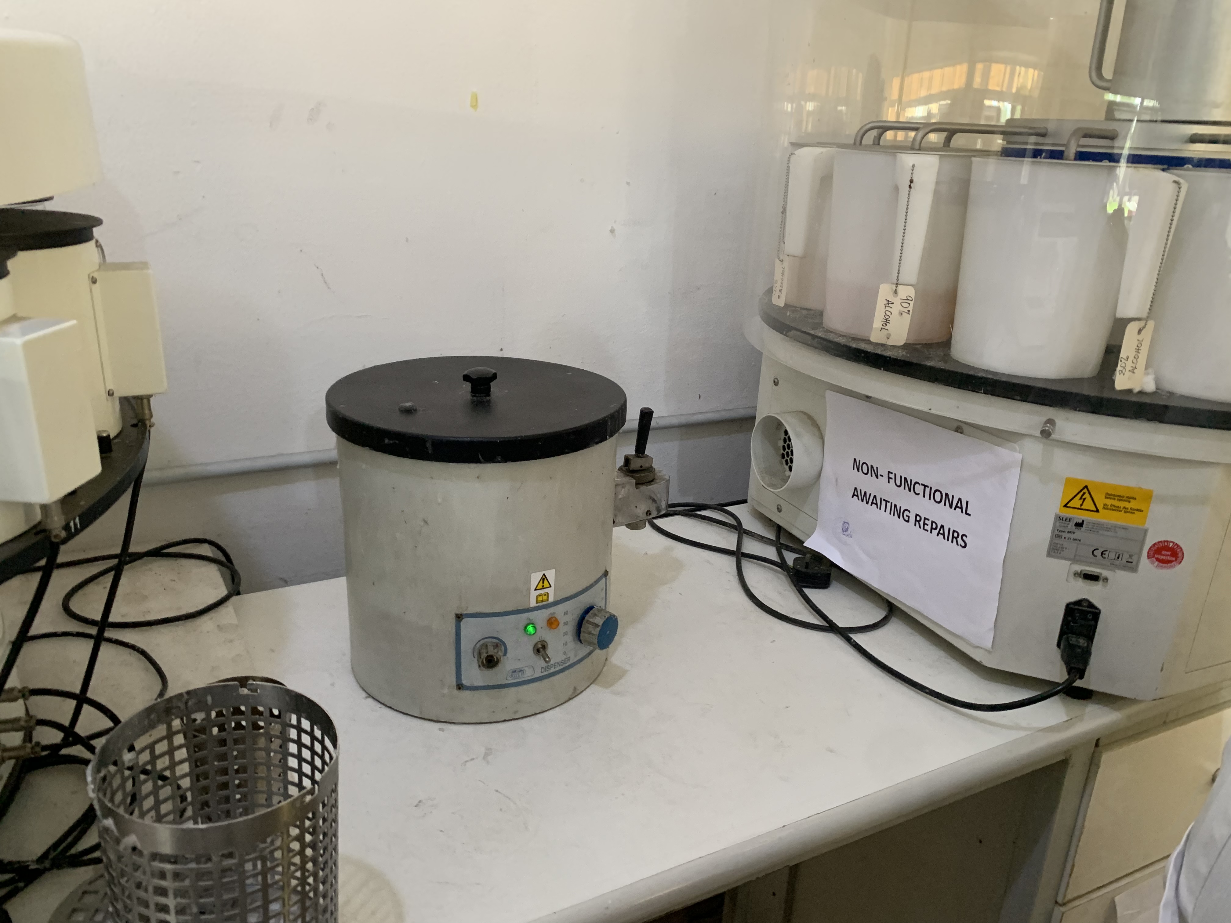
Infiltration container
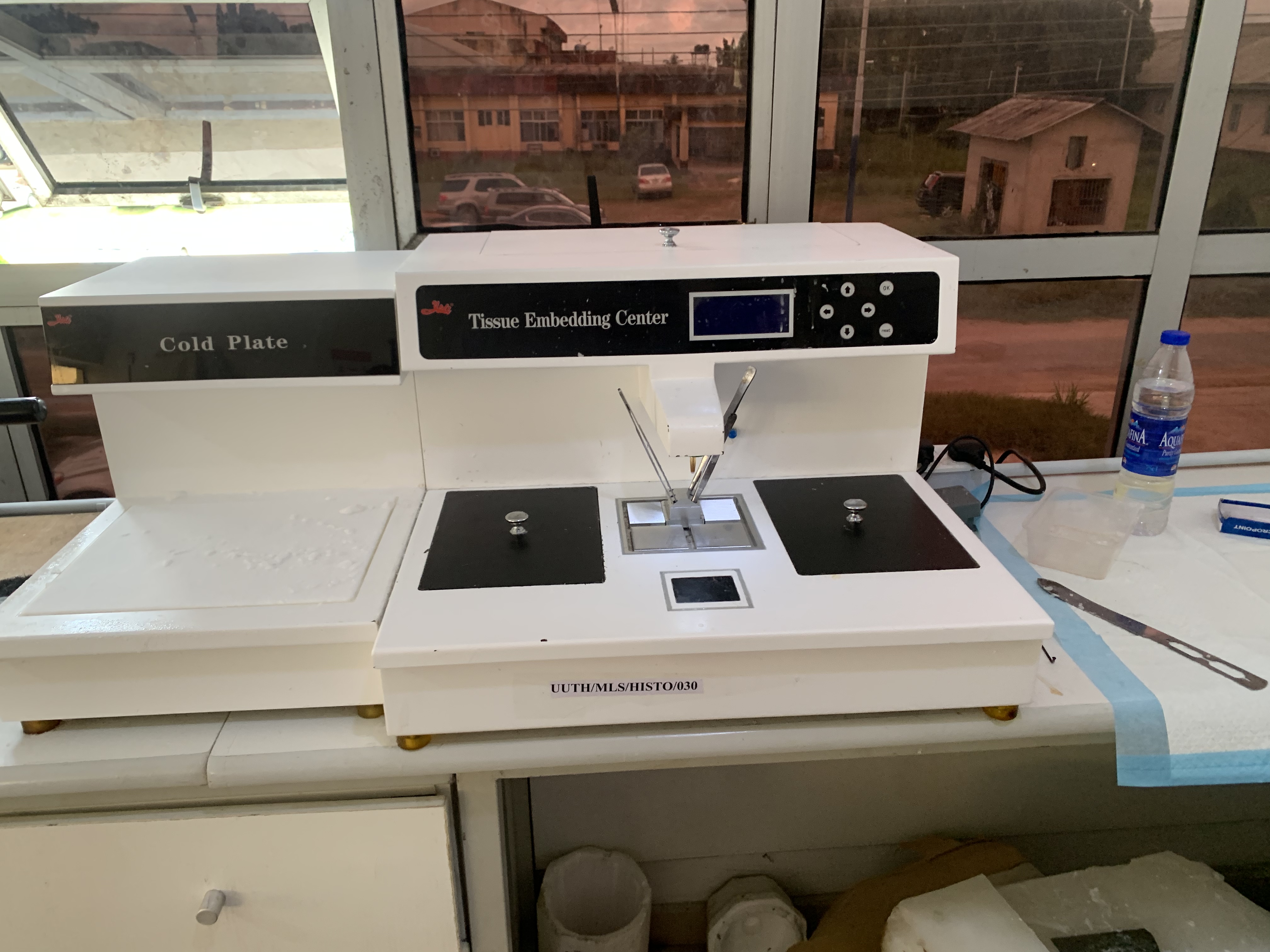
Embedding machine
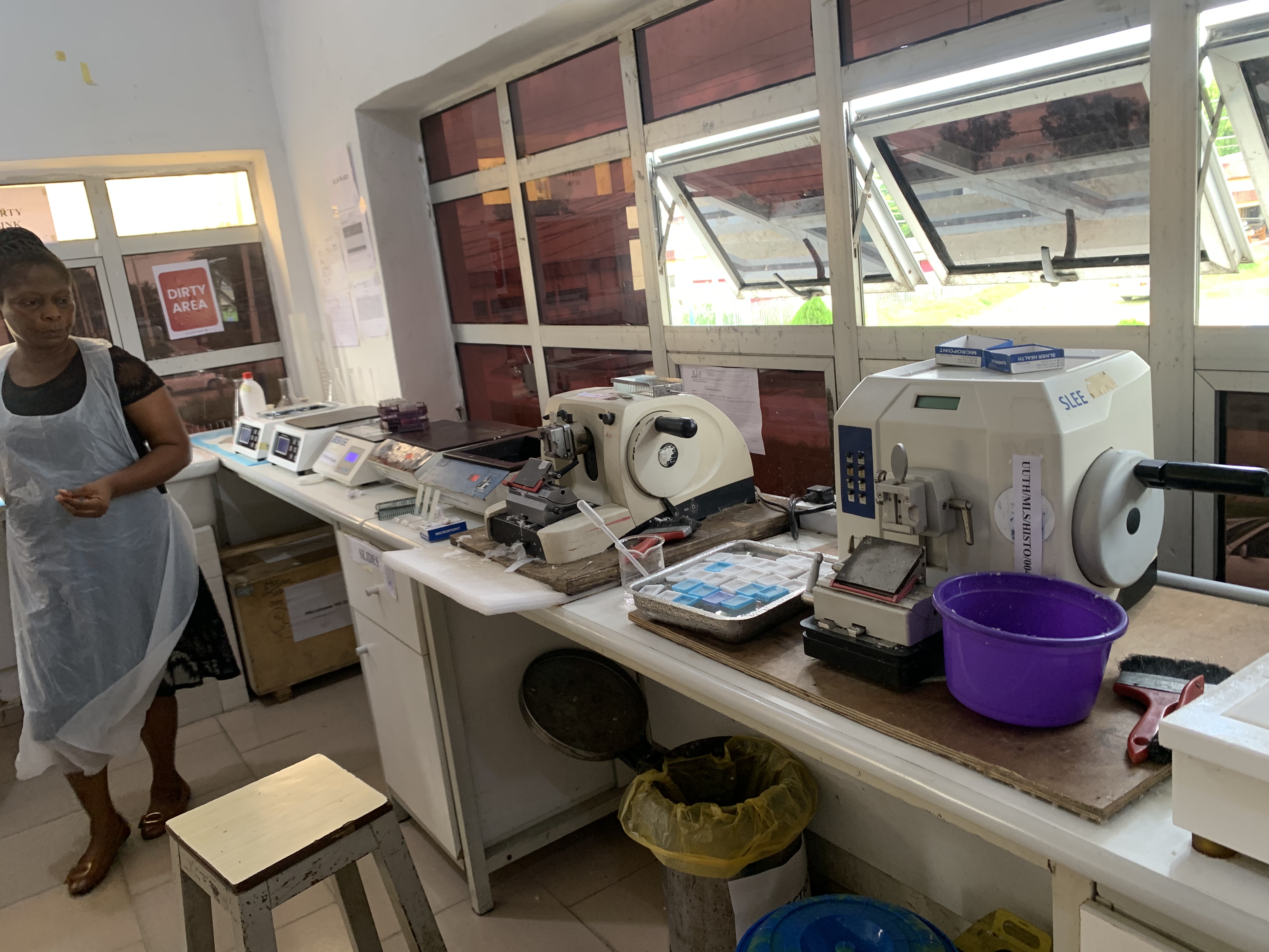
Microtome, water bath and hot plate
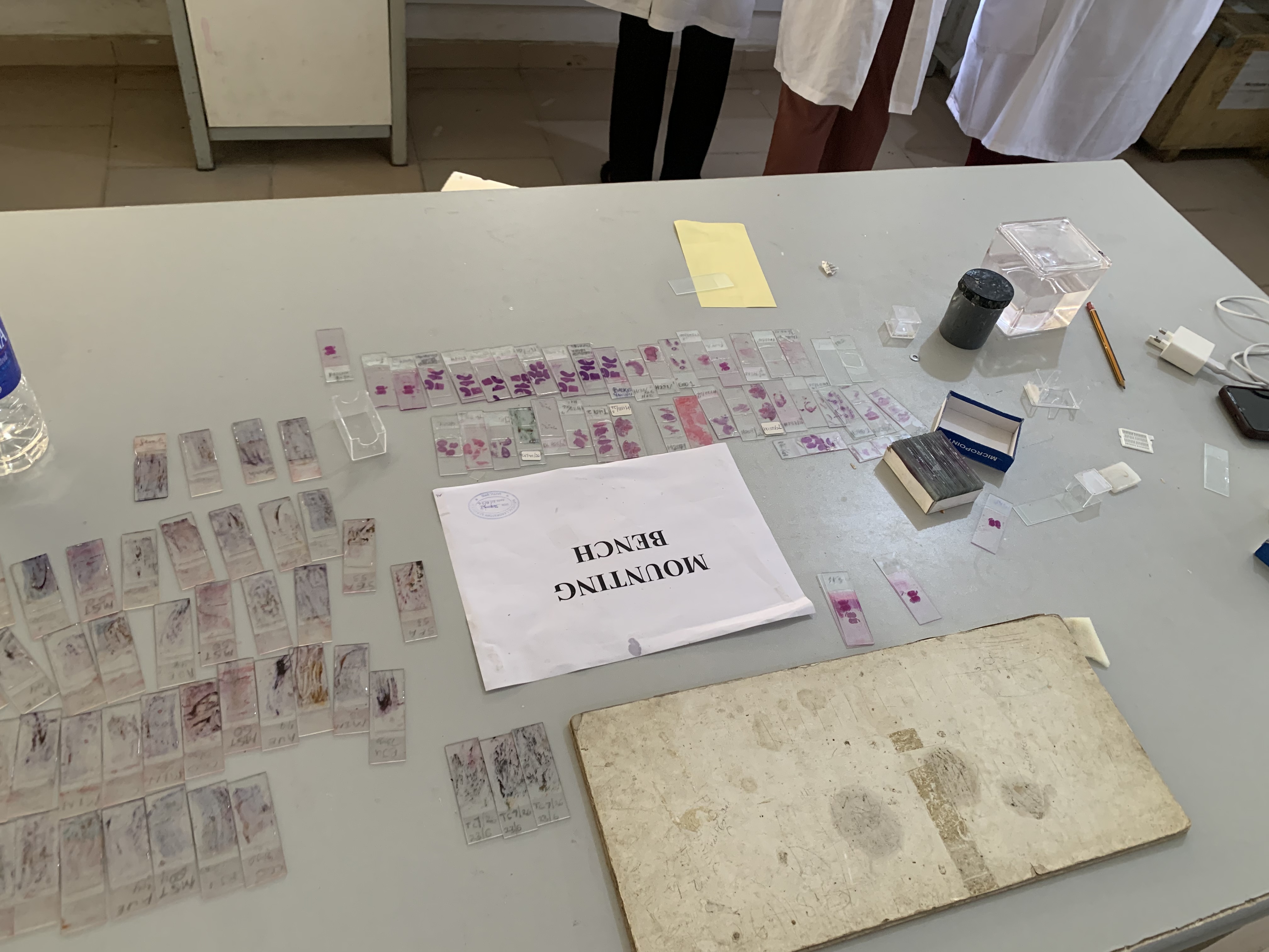
The mounting bench
##LOTS OF HUGS FROM @jennyfav